Xenoturbella bocki
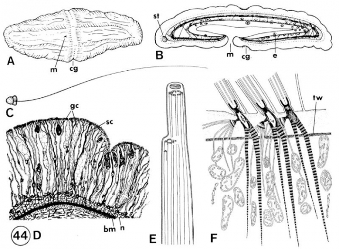
Description A. Ventral view of whole animal. B: Midsaggital section through body. D: spermatozoon. D: Scheme of epidermis.,= E,F: Epidermal cilia. tip (E) and bases showing rootlets (F); anterior=or toward right. (A-C after Ax, 1963 after Westblad, 1949; D after Reisinger, 1960; E and F after Franzen ad Afzelius, 1987). bm, basement membrane; cg, ciliated groove; e, egg; gc, gland cell; m, mouth; n, nerve plexus; sc sensory cell; st statocyst; tw, terminal web.
JPG file - 119.31 kB - 800 x 588 pixels
added on 2017-04-071 057 viewsERMS taxaScan of photo Xenoturbella bocki Westblad, 1949checked Tyler, Seth 2017-04-07
 Comment (0)