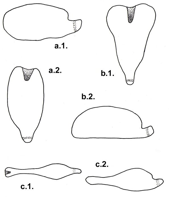
Pourtalesiidae (lateral + ventral)
Description Lateral (1.) and ventral (2.) view of pourtalesiids: a. Pourtalesia jeffreysi Wyville Thomson: The posterior end is strongly rostrate and ringed by a subanal fasciole; length 20-30 mm (after B. David 1983). b. Ceratophysa: The test is very broad, ending in a rostrum; length 98 mm (after B. David 1984). c. Echinosigra phiale: The anterior part of the test is elongated; length approximately 25 mm (after B. David 1984).
JPG file - 139.67 kB - 1 595 x 1 875 pixels
added on 2005-02-231 494 viewsERMS taxaScan of drawing Pourtalesiidae A. Agassiz, 1881checked Kroh, Andreas 2021-02-24Scan of drawing Echinosigra phiale (Thomson, 1873) represented as Echinosigra (Echinosigra) phiale (Thomson, 1873)checked Kroh, Andreas 2021-02-24Scan of drawing Pourtalesia jeffreysi Thomson, 1873checked Kroh, Andreas 2021-02-24From reference Schultz, H. (2006). Sea urchins: A guide to worldwide sha...
Download full size © 2005 Schultz, Heinke
 Comment (0)